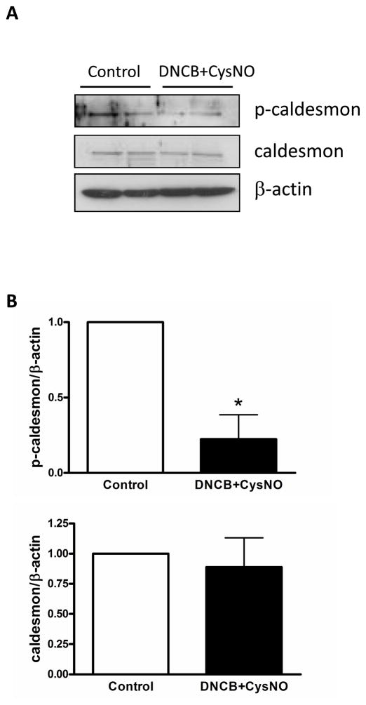
Figure 6.
Phosphorylation of caldesmon was reduced by S-nitrosylation. Isolated mouse aortas were treated with DNCB (4 μM, 1 h) plus CysNO (1.5 mM, 30 min) and then subjected to immunoblot with p-caldesmon, caldesmon, and β-actin antibodies. A: Western blot analysis. B: Bar graphs showing the relative expression of phosphorylation and total proteins after normalization to β-actin expression. Results are presented as mean ± SEM in each experimental group. *, p<0.05 vs. control (n=4).

