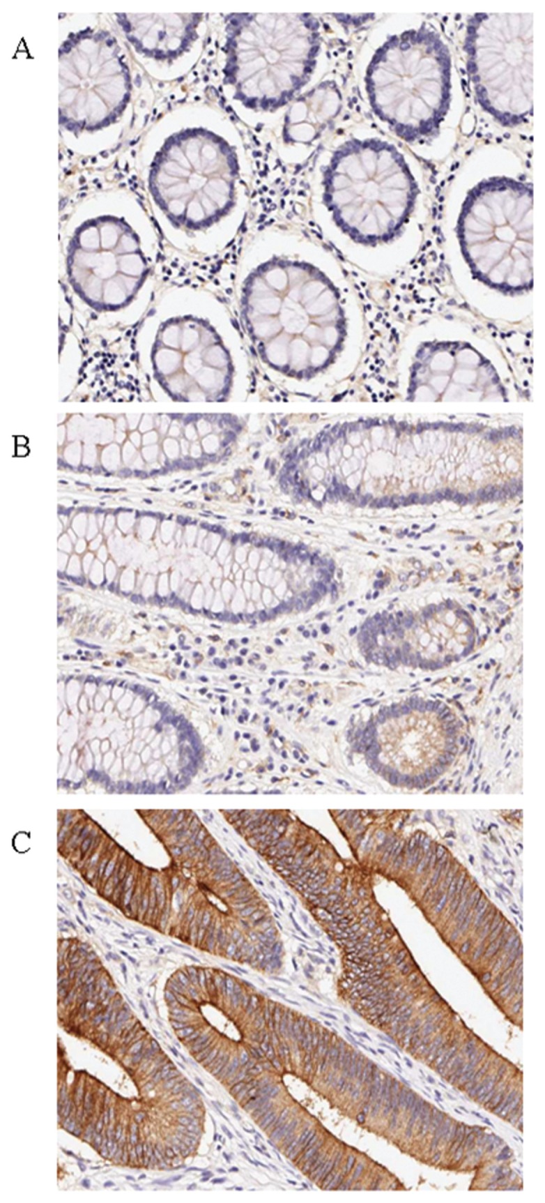
Figure 2.

Immunohistochemical analysis of CRC tissues for PLSCR1 expression (brown color). The paired paraffin-embedded CRC tissue sections were stained with the anti-PLSCR1 antibody. A representative case is presented that contains normal colorectal mucosa, a benign adenomatous polyp and colorectal adenocarcinoma tissue. (A) Low or negative staining of PLSCR1 in normal colorectal mucosa. (B) Upregulated expression of PLSCR1 in benign colorectal polyp tissues. (C) High-level expression of PLSCR1 in colorectal adenocarcinoma tissues. All slides were counterstained with hematoxylin. All photographs are presented at 200× magnification.
