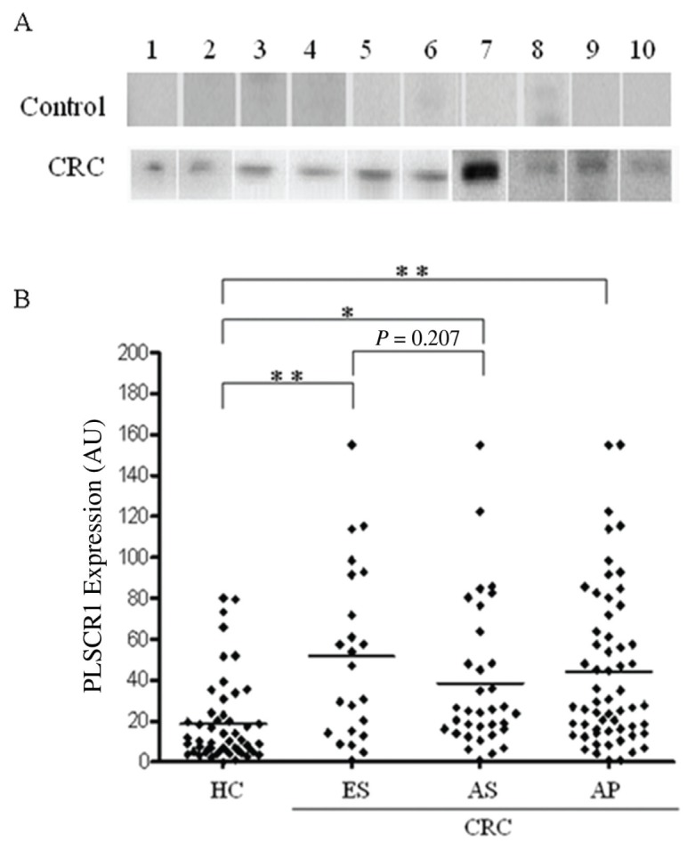
Figure 3.

Western blot analysis of PLSCR1 expression in plasma. (A) Plasma samples (1 μL) from each tested specimen were denatured and loaded onto an SDS-PAGE gel. Each line represents an individual participant. Representative samples from healthy controls with negative or low PLSCR1 plasma levels and CRC patients with elevated PLSCR1 plasma levels are shown. (B) PLSCR1 was elevated in the plasma of CRC patients at different disease stages compared with plasma from healthy controls. HC, healthy controls; ES, early stage (TNM stage I and II); AS, advanced stage (TNM stage III and IV); AP, all patients; the horizontal bar represents the mean value of each group; *P < 0.01, **P < 0.001, Student t test.
