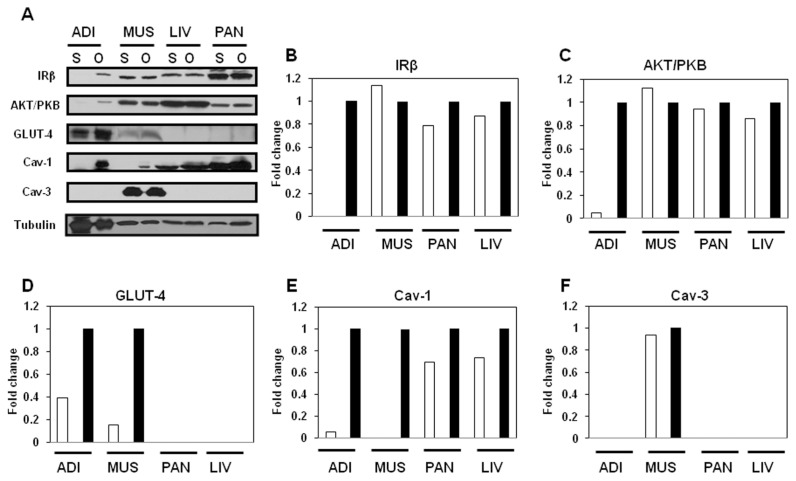
Figure 6.
Expression of insulin-signaling molecules and caveolins in orchidectomized male JYD mice. (A) Insulin-signaling molecules and caveolin proteins were analyzed by Western blot in adipose tissue (Adi), skeletal muscle (Mus), liver (Liv) and pancreas (Pan) of sham-operated (S) or orchidectomized (O) male JYD mice at 5 wks after orchidectomy. Tubulin was used to confirm equal loading. (B–F) Quantitative analysis of Western blots in tissues. The relative fold-change in sham-operated mice (white bars) compared to the control band (orchidectomized mice, black bars) on Western blots was quantified by densitometric analysis.

