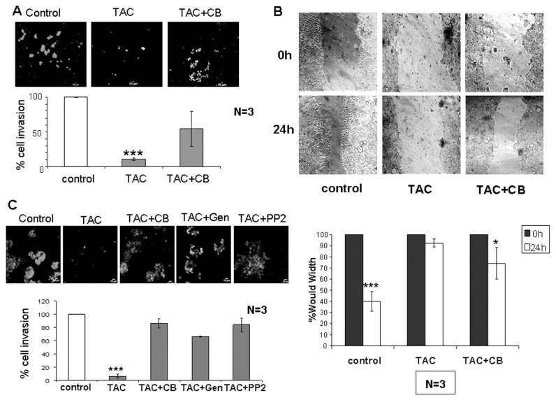
Figure 3.
Effect of testosterone-HSA on Caco2 human colon cancer cell migration and adhesion. (A) Cells were cultured with 10−7 mol/L testosterone-HSA in the presence or absence of 10−6 mol/L cytochalasin B on the Matrigel-coated upper compartment of Tran-swell culture chambers, provided with an 8-μm pore-size polycarbonate filter, according to the manufacturer’s instructions. Matrigel was removed by scraping 24 h later, and invaded cells, attached to the lower surface of the filter, were stained with DAPI. The slices were imaged under the microscope and the number of cells in 10 random fields was counted. Bars represent the % of cell invasion in control and treated cells; (n = 3), ***P < 0.001. (B) Wound healing assay of Caco2 cells. Following 24-h culture the confluent monolayer was scratched with a pipette tip to create a cell-free area. Testosterone-HSA 10−7 mol/L was added, and wound closure was documented by microphotography of the same region after 24 h. Bars represent the width of the wound in control and treated cells; *P < 0.05; ***P < 0.001. (C) Cells in the presence or not of 50 μmol/L genistein and PP2 were cultured with 10−7 mol/L testosterone-HSA in the absence or presence of 10−6 mol/L cytochalasin B on the Transwell culture chambers, provided with an 8-μm pore-size polycarbonate filter, according to the manufacturer’s instructions. After 24 h, invaded cells, attached to the lower surface of the filter, were stained with DAPI. The slices were imaged under the microscope, and the number of cells in 10 random fields was counted. Bars represent the % of cell invasion in control and treated cells; (n = 3), ***P < 0.001.

