Figure 7.
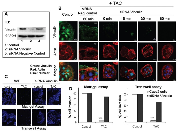
Effect of testosterone-HSA on Caco2 cells silenced with vinculin siRNA (A) Caco2 cells transfected with vinculin siRNA or a negative control siRNA were lysed, and equal amounts of total lysates were immunoblotted (IB) with a specific antibody against vinculin and GAPDH (glyceraldehyde-3-phosphate dehydrogenase). The immunoblots were analyzed by densitometry. (B) Confocal laser-scanning microscopic analysis of vinculin and actin in mAR-activated Caco2 cells silenced either with vinculin siRNA or a negative control siRNA. Transfected cells treated with 10−7 mol/L testosterone-HSA for different time periods were cultured in coverslips, fixed and stained with mouse antivinculin, anti–mouse-FITC as secondary antibody, DRAQ5™ for nuclei staining and rhodamine-phalloidin for filamentous actin. Magnification 100×. (C) Representative motility experiment of Caco2 cells transfected or not with vinculin siRNA. Cells were cultured in the presence of 10−7 mol/L testosterone-HSA on the Matrigel-coated upper compartment or Transwell culture chambers, provided with an 8-μm pore-size polycarbonate filter, according to the manufacturer’s instructions. After 24 h, invaded cells, attached to the lower surface of the filter, were stained with DAPI. (D) Quantification of the motility experiments shown in (C) for Caco2 cells silenced with vinculin siRNA and cultured in the presence of 10−7 mol/L testosterone-HSA as described in Figure 3A and C.

