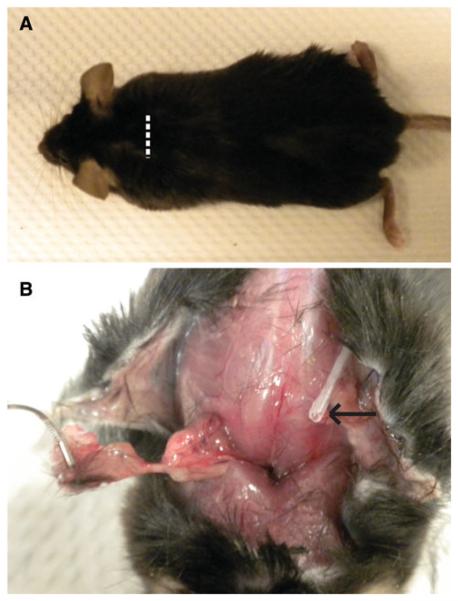
Fig. 1.
a Representative photographs of the mouse 25 days after IAS construct implantation. The white dotted line represents the area of the original incision, which is now healed with hair fully grown over. b The IAS construct (held in forceps) being harvested. The growth factor delivery catheter tip is shown by the black arrow. The tissue in the implantation bed appears healthy without evidence of infection or rejection

