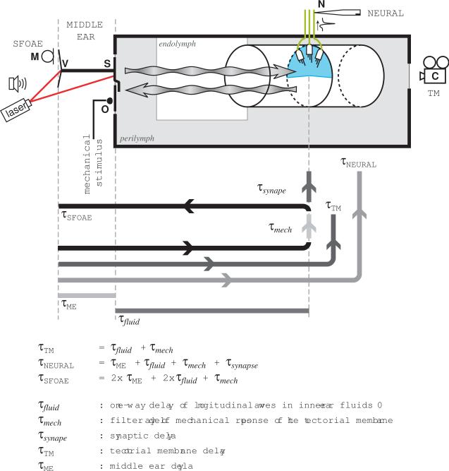
Figure 8.
Diagram illustrating the different recording sites and associated delays for the frog basilar papilla (BP). The stapes (S) connects to the otic capsule (grey rectangle) via the oval window, which also holds the operculum (O). Mechanical vibrations propagate through the inner ear fluids as longitudinal pressure waves (wavy arrows). Within the endolymphatic space (white rectangle), a rigid tubular structure contains the BP sensory epithelium. This is composed of hair cells in the wall of the tube, which are covered by the semicircular tectorial membrane (blue). Hair cells in the BP are only connected by afferent nerve fibers (green lines). The middle ear group delays from Fig. 2 were obtained by comparing the phase of the tympanic membrane velocity (V) with that of the microphone signal (M), or the stapes footplate velocity. For the tectorial membrane group delays (asterisk in Fig. 6), the phase of the mechanical stimulus of the piezo-electric stimulator on the operculum was compared to that of the tectorial membrane, as determined from movies obtained with a high-speed camera (C) focussed through the round window. Neural delays (closed symbols in Fig. 6) were calculated by comparing the acoustic stimulus (M) with the spike times of intracellular recordings from auditory fibers in the auditory nerve (N). The group delays for the SFOAE (circles in Fig. 6) were calculated by comparing the phase of the emissions with those of the stimulus tones, both calculated from the microphone signal (M). Note that τ-values with capital subscripts correspond to measured quantities.

