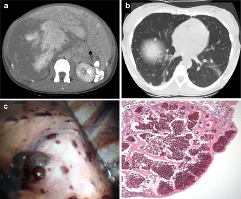
Fig. 1.
a Initial abdominal CT at presentation in 2003 showing the largest lesion (18.1 × 15.9 cm) measured at level of the right portal vein and the second largest lesion was 4.3 × 3.5 cm. b Initial chest CT in 2003 revealing innumerable pulmonary nodules. c Video-assisted thoracoscopic surgery (VATS). Diffuse hemorrhagic, purple/red, raised nodules are seen on the surface of the lung. Biopsy of these lesions revealed benign cavernous hemangiomas. d Photomicrograph of an H&E-stained section of cavernous hemangioma in a lung biopsy specimen (40×). The lung lesions were small and well-circumscribed with benign endothelial cells and thin vessel walls and septa composed predominantly of fibrous tissue.

