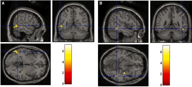
Fig. 3.
BOLD in the left middle temporal gyrus and the right subgyral temporal lobe. Group average activation data for the association of HSP mean with the minor less the major change condition in the (A) left middle temporal lobe and the (B) right subgyral temporal lobe. Lighter color corresponds to greater activation. MNI co-ordinates for the center of the left (second peak) and right activation clusters were −56, −54 and −4 and 48, −46, −12, respectively.

