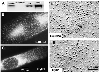
Figure 1.
RyR1 carrying the E4032A mutation is properly expressed in 1B5 myotubes. 1B5 cells grown in collagen-coated 35-mm dishes were infected with either RyR1 or E4032A cDNA-containing herpes simplex virus amplicon virions at 3 × 10⁵ IU/ml. (A) Western blot of 1B5 myotube preparations expressing E4032A-RyR1 (lane 1, 20 μg), 1B5 null myotubes not transduced with cDNA (lane 2, 20 μg), and 1B5 myotubes expressing wtRyR1 (lane 4, 20 μg). Lane 3 contains 0.5 μg of rabbit junctional sarcoplasmic reticulum as a positive control. (B) The intracellular distribution of E4032A expressed in 1B5 myotubes was examined by using immunocytochemistry as described in Materials and Methods. E4032A was properly targeted to junctional domains at the fiber periphery as indicated by the punctate appearance of the immunolabeling pattern which was indistinguishable from the pattern obtained when wtRyR1 was expressed in 1B5 cells (C). (D and E) E4032A- and wtRyR1-expressing 1B5 myotubes were examined for DHPR tetrad formation by freeze–fracture electron microscopy. Both RyRs induced tetrad formation in the plasmalemma of 1B5 cells as indicated (arrows). The tetrads were similar in appearance and indicate the formation of a stereospecific link between four DHPRs and the four subunits of the RyR.

