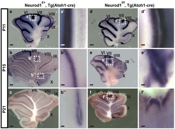
Fig. 13.
Spatial and temporal expression of Barhl1 changes in absence of Neurod1 in cerebellum, as shown by in situ hybridization of Barhl1. a, a’ Barhl1 was expressed in EGL in all lobules and in IGL, was highly expressed in anterior lobules I-1/2VI, absent in lobules VII and VIII, and low in lobules IX and X in P11 heterozygous cerebellum. b-b” By P15, Barhl1 expression was downregulated in IGL and EGL in heterozygous cerebellum. c By P21, Barhl1 expression was diminished in heterozygous mice. d, d’ In P11 mutant cerebellum, IGL was almost devoid of Barhl1 expression, except for some in lobule X; EGL showed expanded Barhl1 expression. e, e’ Barhl1 expression persisted and expanded in EGL predominantly in central lobules in mutant P15 cerebellum, instead of being expressed in IGL. f, f’ Barhl1 abnormally continued to be expressed in EGL of central lobules in P21 mutant cerebellum. The boxed areas in a-f are shown at higher magnification in a’-f’. Bars 250 μm (a-f), 50 μm (a’-f’, b”)

