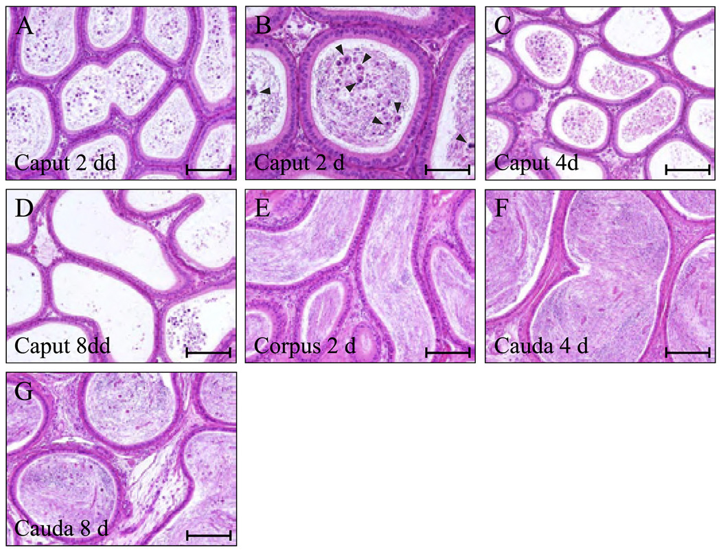
FIG. 8.
A study to examine the effects of adjudin in the epididymis. A single oral dose of adjudin at 50 mg/kg b.wt. was administered to adult rats. On days 2, 4, and 8, epididymides were removed, dissected into three segments [caput (initial segment), corpus (middle segment), and cauda (final segment)] and processed for histological analysis after hematoxylin & eosin staining using paraffin sections. By days 2 and 4 after treatment, immature germ cells such as spermatocytes and early spermatids (see arrowheads in B) were detected in the caput (A–C), but by day 8, these cells had depleted this segment of the epididymis (D). On days 2 and 4, normal epididymal spermatozoa were found in the corpus (E) and cauda (F), and apparently their function was unaffected by adjudin, because all animals remained fertile until the epididymis became devoid of all spermatozoa by ~30 days after treatment (Cheng et al., 2005a). By day 8, however, immature germ cells were seen in the cauda mixed together with normal epididymal spermatozoa (G). In addition, adjudin did not affect adhesion between epididymal epithelial cells. Bars, A and C to G, 120 µm; B, 60 µm.

