Figure 3.
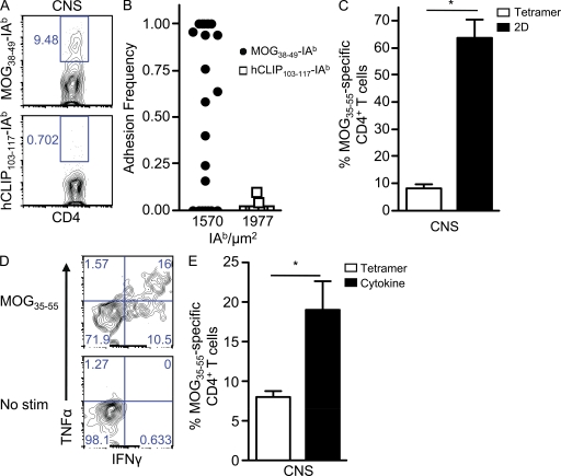
Dominance of proinflammatory low affinity myelin-reactive CD4+ T cells during EAE. (A and B) Representative tetramer (A) and 2D binding (B) of CNS-infiltrating CD4+ T cells to MOG38–49-IAb and hCLIP103–117-IAb. (C) The mean frequency ± SEM of MOG35–55-specific binding by tetramer and 2D analysis was based on three experiments performed in parallel, and CNS tissue was pooled together from 6–10 mice per experiment (*, P = 0.004). (D) Representative frequency of cytokine-producing (IFN-γ and TNF) CD4+ T cells isolated from the CNS during acute EAE after stimulation with MOG35–55 or no peptide (no stimulation [stim]). (E) The mean percentage ± SEM of CNS-infiltrating MOG35–55 CD4+ T cells was compared in parallel by MOG38–49-IAb tetramer and the percentage producing cytokine (IFN-γ and TNF) upon stimulation with MOG35–55 (no stimulation background subtracted) from two independent experiments (CNS tissue pooled from 12–24 mice; *, P = 0.04).

