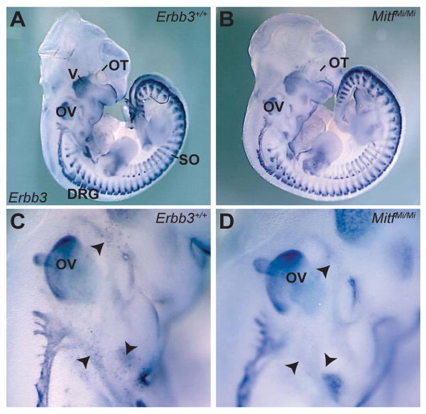
Figure 1. Erbb3is expressed in MC precursors during embryonic development.
Lateral view of Erbb3 whole-mount in situ hybridization in wild-type (A, C) and MitfMi/Mi (B, D) embryos at E11.5. In both wild-type embryos and MitfMi/Mi mutants, Erbb3 expression is observed in the cranial ganglia and nerves, DRG and somites. In wild-type embryos (C), a population of Erbb3-expressing cells (black arrowheads) is detected in the vicinity of the otic vesicle, the location where Mbs are normally found. These Erbb3-expressing cells were absent in Mb-deficient mutants (D), indicating that these cells are Mbs. Labeled structures are otocyst (OT) trigeminal ganglia (V), dorsal root ganglia (DRG), otic vesicle (OV) and somites (SO).

