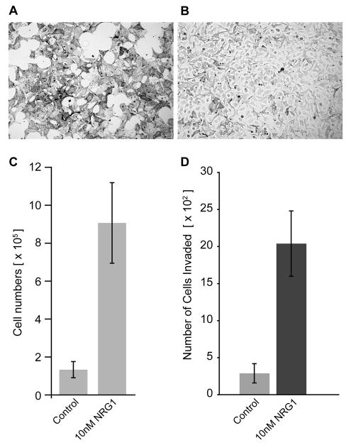
Figure 4. NRG1 increases proliferation and invasion of melan-Ink4a cells.
Bright filed images of melan-Ink4a cells (A, B) in media lacking TPA and CT (A) and in media lacking TPA and CT but with 10nM NRG1 added (B). (C) Cell doublings in the absence of TPA and CT (control) and in media containing NRG1 without TPA and CT (10 nM NRG1). Cells were plated at equal numbers (2.5 × 10⁵) and total numbers of cells were counted four days after the indicated treatments using hemocytometer. While melan-Ink4a grown in the absence of TPA/CT stopped proliferating, addition of NRG1 increased number of Melan-Ink4a cells. D) Increased invasion in the presence of NRG1. Control and NRG1-treated Melan-Ink4a cells were grown in Boyden chambers and assessed for their ability to invade through basement membrane matrix. For quantification, stained cells were photographed on Zeiss Axiovert 135 microscope and cells were counted in at least 16 random fields. Columns represent the absolute number of cells that invaded the matrix 48 hours after seeding (n=4).

