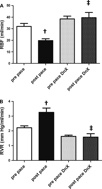
Fig. 3.
Renal hemodynamics (protocol 1). A: RBF was measured via flow probe around the left renal artery in 5 intact and 5 DnX animals before and after 3 wk of continuous pacing The value for 1 animal is the average of a 10-min recording on 3 separate days. †P < 0.05 vs. respective prepace group. ‡P < 0.05 vs. postpace; n = 5. B: RVR in 5 intact and 5 DnX animals before and after the pacing protocol. The value for 1 animal is the average of a 10-min recording on 3 separate days. †P < 0.05 vs. respective prepace group. ‡P < 0.05 vs. postpace; n = 5.

