Fig. 5.
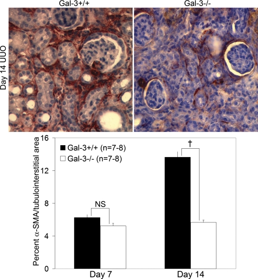
Interstitial α-SMA+ myofibroblast numbers were significantly lower in Gal-3-deficient mice compared with wild-type mice at day 14 after UUO despite an increase in fibrosis severity. Representative α-SMA-stained immunohistochemical photomicrographs (×400) are shown at top and the graph at the bottom summarizes the quantitative results of α-SMA interstitial staining. Results are expressed as means ± SE. †P < 0.01, wild-type vs. Gal-3-deficient.

