Fig. 7.
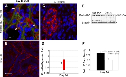
Attenuated expression of Endo180 in Gal-3-deficient mice. A: dual stained confocal microscopy of collagen I (green) and F4/80 (red). Arrows highlight F4/80+ macrophages integrated within collagen matrix. B: photomicrograph illustrates the diffuse interstitial infiltrate of Endo180+ cells (red) at day 14 after UUO. C: photomicrograph illustrates interstitial expression of α2 integrin, a potential binding partner for Gal-3. D: boxplot summarizes analysis of relative Endo180 mRNA expression normalized to both GAPDH and 18S, Gal-3-deficient to wild-type, measured by semiquantitative real-time qPCR. E: Endo180 Western blot of total kidney protein shows decreased expression levels and graph (F) summarizes results of normalized Endo180 to β-actin levels at day 14. Results are expressed as means ± SE. ‡P < 0.05, wild-type vs. Gal-3-deficient. Bar = 10 μm (A); bar = 20 μm (B, C).

