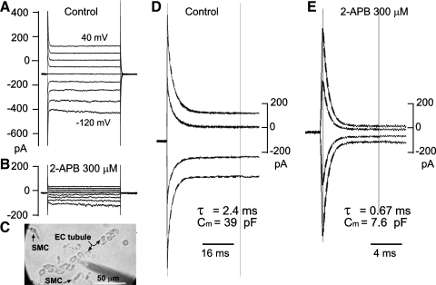
Fig. 7.
2-APB suppressed the gap junction coupling with little other membrane effects in ECs tubules from the SMA. A and B: current traces elicited by 500-ms step commands in the absence (A) and presence (B) of 2-APB from a cell in a tubule of ECs (C; the electrode pipette points to a cell other than that in A). D and E: representation of the initial part of A and B, respectively, but with traces of −100-, −60-, −40-, and 20-mV steps removed and the time scale expanded for clarity. Note that single-term exponential function fit well with the transients in both the absence and presence of 2-APB. Rinput increased from 334 MΩ to 1.7 GΩ, suggesting that 300 μM 2-APB completely isolated the recorded cell in the control condition, under which the cell appeared tightly coupled to at least four other cells. The EC showed an obvious inward, but not outward, rectification in the voltage range tested.

