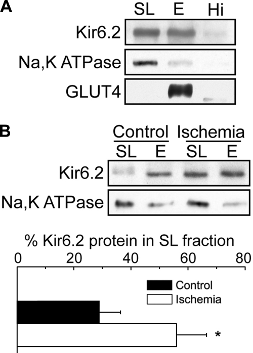
Fig. 5.
Increase in cardiac myocyte sarcolemmal KATP channels following global ischemia. A: cell fractionation was performed on rat ventricular tissue from nonischemic and ischemic rats using Optiprep gradient centrifugation. The sarcolemmal fraction (SL = 0/5% interface), endosomal fraction (E = 5/15% interface), and a loose pellet representing the highest density material (Hi) were subjected to Western blot analysis. B: representative Western blot of the SL and E fractions of membranes prepared from nonischemic control and ischemic rat hearts. The results from 3 experiments were quantified and plotted below as means ± SE. *P < 0.05. Figure panels have been adjusted for brightness and contrast.

