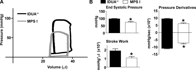
Fig. 1.
Baseline hemodynamic function. A: representative raw pressure-volume loops of lysosomal hydrolase α-l-iduronidase (IDUA+/−; black) and mucopolysaccharidosis type I (MPS-I; gray) mice. B: mean hemodynamic data showing differences in cardiac performance at baseline. *P < 0.05 by t-test. Wild-type (WT), n = 7; MPS-I, n = 8.

