Fig. 1.
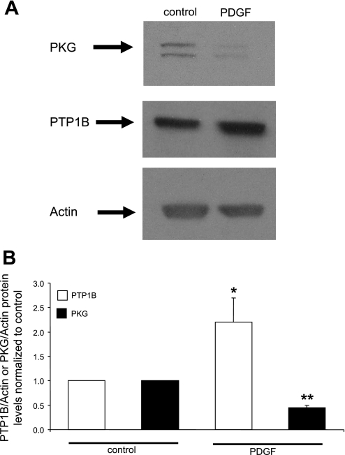
PDGF induces a suppression of PKG levels together with an upregulation of protein tyrosine phosphatase 1B (PTP1B) levels. Aortic smooth muscle cells were cultured in serum-free medium for 24 h followed by treatment with 20 ng/ml PDGF in serum-free medium for 24 h. A: Western blots from a representative experiment. Duplicate bands in the PKG blot represent α- and β-isoforms. B: summary of results (mean ± SE) from 3 experiments, normalized to α-actin levels. *P < 0.05, PDGF + PTP1B compared with control; **P < 0.05, PDGF + PKG compared with control.

