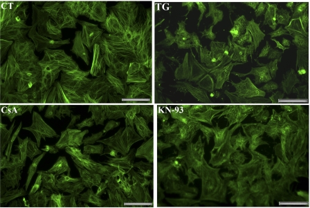
Fig. 1.
Fluorescence microscopy of cultured neonatal rat cardiac myocytes. Cardiac myocytes were prepared as described in methods and maintained in serum-free medium. As indicated, the culture medium was supplemented with 10 nM thapsigargin (TG), 200 nM cyclosporin (CsA), or 5 μM KN-93 (added 24 h after seeding). The myocytes were then fixed after 3 days, permeabilized, stained with Alexa Fluor 488-phalloidin, and viewed with a fluorescence microscope (×20 magnification). The images shown are representative of numerous observations of independent preparations, and the magnification bar corresponds to 50 μm.

