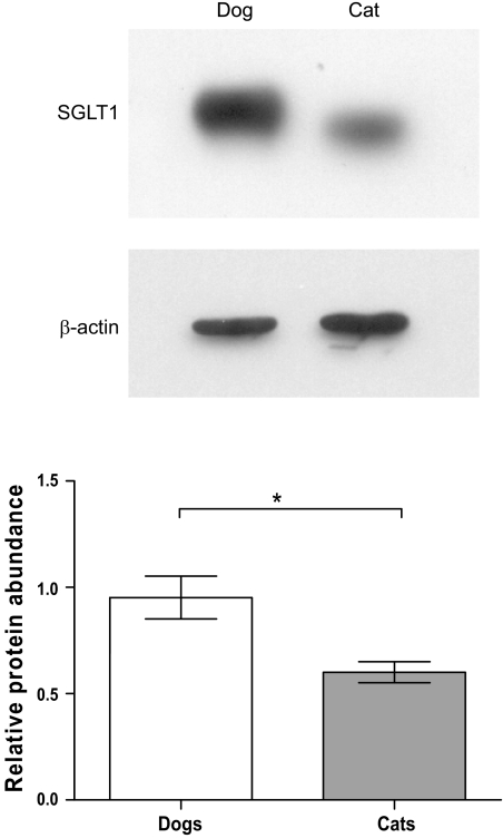
Fig. 4.
Representative Western blots (top), showing SGLT1 and β-actin protein abundance in BBMV prepared from the middle intestine of dogs and cats. Densitometric scans of Western blots normalized to β-actin are presented as histograms (bottom); dogs (open bar) and cats (gray bar). Results are shown as mean with SD. Significant difference *P < 0.05.

