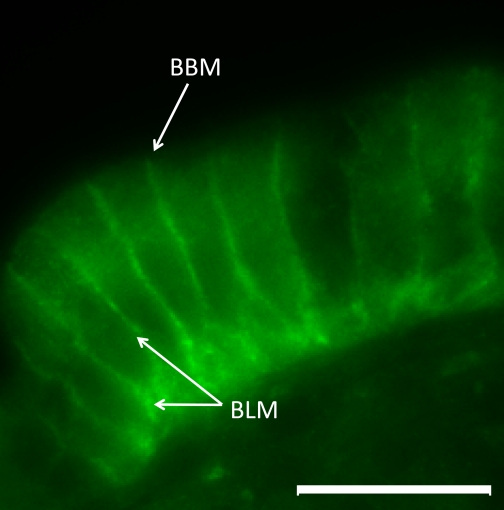
Fig. 7.
A representative image showing immunofluorescent localization of GLUT2 in the intestinal mucosa of dogs or cats. Signal for GLUT2 is observed along the basolateral membrane of enterocytes, with no detectable signal at the brush border membrane. ×1,000 magnification, scale bar = 10 μm. BBM, brush border membrane; BLM, basolateral membrane.

