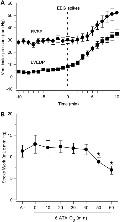
Fig. 5.
Ventricular pressures and left ventricular function in anesthetized rats in 6 ATA O2. Mean values of cardiac parameters are shown for 8 anesthetized animals that demonstrated EEG spikes during 60-min exposure. A: RVSP and LVEDP are shown for 10 min before and after the onset of EEG spikes (time 0). B: stroke work (stroke volume × mean arterial pressure), an index of left ventricular function, was calculated every 10 min. Left ventricular function (stroke work) diminished significantly in these rats, which exhibited EEG spikes. Values are means ± SE. *P < 0.05 vs. air.

