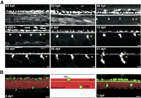
Fig. 1.
Time course of Rohon-Beard (R-B) neuron degeneration in Isl2b:EGFP transgenic zebrafish. A: enhanced green fluoresent protein (EGFP) expression driven by Isl2b promoter facilitates observation of R-B neurons degeneration concurrent with development of dorsal root ganglia (DRG, arrow heads). All images of the spinal cord are maximum intensity projections of stacks acquired in the lateral plane using confocal microscopy over a period from 24 h postfertilization (hpf) to 31 days postfertilization (dpf). B: merged confocal images (left and right) and schematic representation (middle) of the double-labeled transgenic line (Isl2b:EGFP/HuC:mCherry) imaged in the lateral plane. The contrast and transparency of the images were adjusted to emphasize EGFP-labeled sensory neurons. The boundaries of the spinal cord are delineated by the mCherry expression. Note the anterior and posterior axonal projections and location of EGFP-labeled sensory neurons over the dorsal longitudinal fasciculus (DLF) in the double-labeled embryo spinal cord. All scale bars represent 20 μm and all images are oriented with anterior to the left and dorsal to the top.

