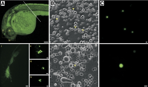
Fig. 2.
Isolation of single R-B neurons from Isl2b:EGFP transgenic zebrafish embryos. A: at 24 hpf, several clusters of neurons (labeled i–iv) in the cranial region displayed intense EGFP expression. - - -, where embryos were transected prior to enzymatic and mechanical dissociation of the trunk region. The fluorescence in the yolk sac was auto-fluorescence. B and C: phase-contrast (B) and fluorescent (C) photomicrographs of acutely dissociated R-B neurons (▶) from 24 hpf Isl2b:EGFP embryos. Scale bar in A (top) represents 1 mm. The remainders of the scale represent 10 μm.

