Fig. 4.
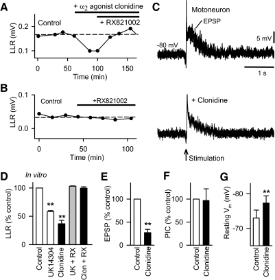
Activation of the α2 adrenergic receptor does not directly affect the Ca PIC but instead inhibits EPSPs. A: amplitude of LLR recorded in the isolated in vitro spinal cord of chronic spinal rat and measured repeatedly over time (LLR quantified 0.5–4 s post stimulus, as in Fig. 2). Control values are shown on left, followed by activation of α2 adrenergic receptors with the agonist clonidine (top bar 0.1 μM) and subsequent application of the α2 receptor antagonist RX821002 (0.3 μM; bottom bar). B: same format as A with application of RX821002 alone (0.5 μM). C: intracellular motoneuron recording of long-latency polysynaptic EPSP (abbreviated EPSP) evoked by dorsal root stimulation (0.1 ms at 3 × T) of chronic spinal rat (quantified at 200 ms post stimulus) during hyperpolarizing bias current before (top plot) and after blocking the α2 receptor with bath application of clonidine (0.3 μM, bottom plot). D: normalized group mean for LLR in chronic spinal rats in vitro with application of the α2 receptor agonists UK14304 (0.03 μM; n = 8), and clonidine (0.1 μM; n = 5), and application α2 antagonist RX821002 (0.3 μM) after UK14304 (abbreviated UK14 + RX; n = 8) and after clonidine (abbreviated Clon + RX; n = 8). Normalized group mean of intracellulary recorded polysynaptic EPSP (E) evoked by 3 × T dorsal root stimulation, PIC (F), and resting membrane potential (Vm; G) before and after application of clonidine in chronic spinal rats (0.1–1 μM; n = 5). *P < 0.05, **P < 0.01. Error bars, SE.

