Fig. 2.
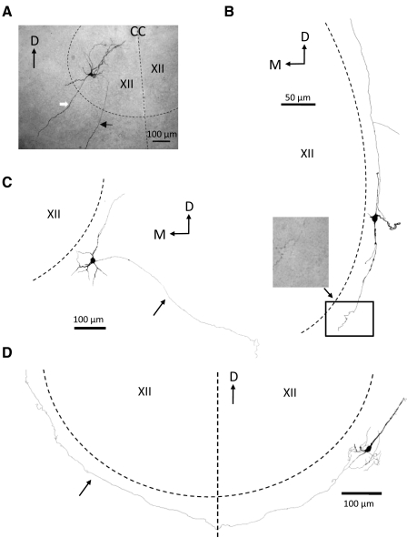
Examples of several morphological types of biocytin-filled mouse brain stem neurons examined in this study. A: photomicrograph of a HM filled with biocytin through a whole cell intracellular recording electrode (see methods). Slice (250 μm thick, nonrhythmic) was processed for biocytin with ABC-peroxidase as a whole mount. The cell body was located in the ventrolateral part of the XII nucleus (therefore likely a genioglossus motoneuron). The approximate boundary of the nucleus and the midline of the brain stem are indicated by the dashed black lines. The black arrow points toward the axon that, after leaving the nucleus, ran ventrolaterally in the slice toward the ventromedial surface of the slice. Note the absence of axon collaterals. The white arrow marks a large dendritic process that extends well beyond the boundary of the XII nucleus. B: camera lucida reconstruction of a multipolar biocytin-filled GFP+ cell located in the NR. The processes of this cell ran parallel to the boundary of the XII nucleus (dashed line marks approximate boundary location). Note the many axonal swellings—presumed synaptic contacts—in the photomicrograph of the terminal axonal arborization of this cell corresponding to the rectangular area in the drawing. C: camera lucida reconstruction of a multi-polar GFP+ cell located in the NR. Arrow points towards the axon which ran in a ventro-lateral direction in the slice away from the XII nucleus. D: camera lucida reconstruction of a multipolar GFP+ cell located in the NR. Arrow points to the cell axon which ran along the border of the XII nucleus (dashed line), crossed the midline (vertical dashed line), and ran along the border of the contralateral XII nucleus. D, dorsal; M, medial; CC, central canal; XII, hypoglossal nucleus.

