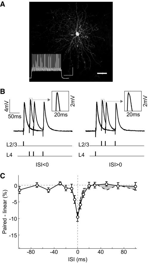
Fig. 7.
Integration window in fast-spiking cells. A: fluorescence image of a neurobiotin-filled nonpyramidal neuron; scale bar, 50 μm. Inset: fast spiking pattern of this cell evoked by current injection (580 pA, 500 ms); scales, 30 mV, 100 ms. B: paired PSPs and linear sums at 3 different ISIs (30, 50, and 100 ms) in a fast-spiking cell. Each trace was averaged from 10 trials. C: integration window for fast-spiking cells (n = 17). Error bar, ±SE.

