Fig. 3.
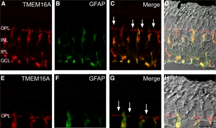
TMEM16A is expressed in both the outer plexiform layer (OPL) and Müller cell bodies, which span the entire retina. A: retinal section immunolabeled for TMEM16A (red). B: Müller cells immunolabeled in a retinal section with glial fibillary acidic protein (GFAP, green). C: merged fluorescent image of TMEM16A and GFAP. D: merged TMEM16A (red) and GFAP (green) immunofluorescence with the differential interference contrast (DIC) brightfield image. Arrows in C indicate labeling of TMEM16A in photoreceptor terminals. E: high-magnification zoom region in the OPL illustrating TMEM16A immunolabeling in the synaptic terminals of photoreceptors (red). F: GFAP labeling of the same high-magnification region (green). G: merged fluorescent high-magnification image of TMEM16A and GFAP. H: merged TMEM16A (red) and GFAP (green) with the differential interference contrast (DIC) brightfield image. Arrows in G indicate labeling of TMEM16A in individual photoreceptor terminals. OPL, outer plexiform layer; INL, inner nuclear layer; IPL, inner plexiform layer; GCL, ganglion cell layer. Scale bars = 10 μm.

