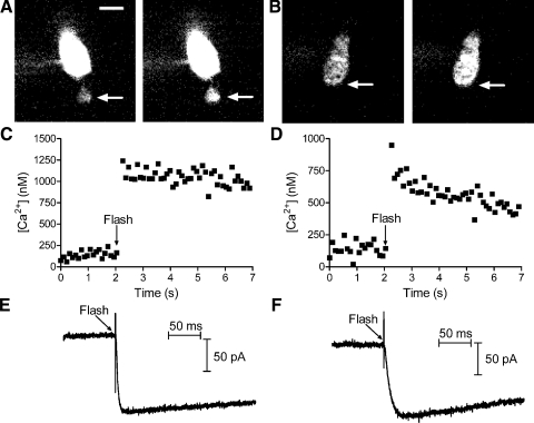
Fig. 6.
Flash photolysis of caged Ca2+ in photoreceptor terminals instantaneously elevated intraterminal [Ca2+] and stimulated inward Cl(Ca) currents. A: grayscale images of Oregon Green BAPTA-6F (OGB-6F) fluorescence in a single confocal section from a rod loaded with the reagent DM-nitrophen. Images were obtained prior to flash photolysis (left) and immediately after flash photolysis (right). Intracellular calcium ion concentration ([Ca2+]i) was measured from a region of interest placed in the synaptic terminal (arrows). Fluorescence is brighter in the soma due to the presence of more dye. B: grayscale images of OGB-6F fluorescence in a single confocal section from a cone before (left) and after (right) flash photolysis of DM-nitrophen. C and D show intraterminal [Ca2+] measured at 60-ms intervals in the rod (C) and cone (D). E and F show inward Cl(Ca) currents evoked in the same rod (E) and cone (F). Cells were voltage-clamped at −77 mV. Ca2+-activated K+ currents were inhibited by Cs+ and tetraethylammonium (TEA) in the pipette solution and reversal potential of chloride (ECl) = −39 mV. Scale bar = 5 μm.

