Figure 7.
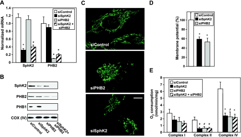
SphK2 and PHB2 regulate HeLa cell mitochondrial function. HeLa cells were transfected with siControl, siSphK2, siPHB2, or both, as indicated. A, B) mRNA levels (A) and protein (B) were determined by quantitative PCR and Western blotting, respectively. C) Mitochondria were stained with anti-cyclophilin D antibody and visualized by confocal microscopy with a ×63 objective. Scale bar = 10 μm. D) Transfected HeLa cells were stained with TMRM, and mitochondrial membrane potential was determined by confocal microscopy with a ×40 objective. Data are expressed as mean ± se percentage of siControl. E) Maximum rates of respiration in the presence of 2 mM ADP were measured polarographically with respiratory complex I, II, and IV substrates. Data are expressed as mean ± se respiration (nmol O2 · min−1 · mg protein−1). Similar results were obtained in 3 additional experiments. *P < 0.05 vs. siControl.

