Figure 4.
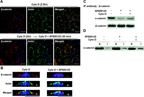
Release of α-catenin from β-catenin is independent of actin polymerization. Human primary keratinocytes were pretreated with actin polymerization inhibitor, cytochalasin D (Cyto D; 2 μM), for 2 h. Next, the cells were treated with either vehicle or SP600125 (10 μM) in the presence of Cyto D for 30 min. A) Confocal images of cells stained with β-catenin (red) and actin phalloidin (green). Nuclei were visualized with Hoechst (blue). View: ×63. Scale bars = 20 μm. B) Lateral images were formed by maximum intensity projection from confocal z-stack images. C) Cell lysates were immunoprecipitated with antibody against β-catenin and subsequently subjected to Western blotting for α-catenin. β-Catenin was used as loading control. D) Soluble (S) and insoluble (I) fractions were separated and subjected to Western blotting for α-catenin.

