FIGURE 2.
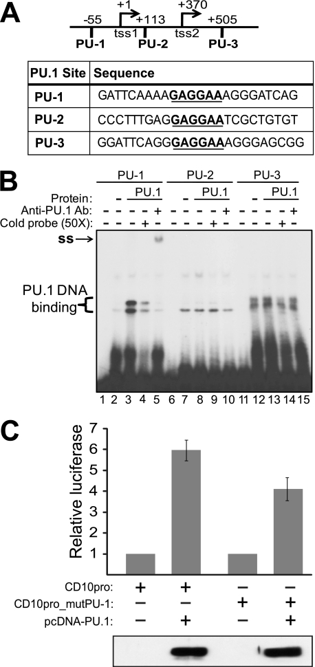
PU.1 binds to a site in the CD10 promoter/enhancer region, which is important for PU.1-induced transactvation of the CD10 promoter. A, the CD10 promoter region has five transcription start sites; the two that are most 5′ are indicated in the figure (tss1 and tss2) (see also Ref. 16). Shown are the three putative PU.1 binding sites (PU-1, PU-2, and PU-3) and the sequences used for EMSA analysis. The PU.1 core sequence is indicated with bold underline. B, nuclear extracts (Protein) from non-transfected A293 cells (−) or A293 cells overexpressing PU.1 (PU.1) were incubated with 32P-labeled DNA probes (PU-1, PU-2, and PU-3). Lanes 1, 6, and 11 contain probe alone (no extract). Where indicated (lanes 4, 9, and 14), a 50-fold excess of cold probe was used to compete the radiolabeled probe. Where indicated (lanes 5, 10, and 15), a PU.1-specific antibody was included to supershift (ss) the protein-DNA complex. C, A293 cells were transfected with pCD10-luc or pCD10mut-luc (with the PU-1 site mutated) and either vector alone (pcDNA) or pcDNA-PU.1; a plasmid encoding β-galactosidase was also included for transfection normalization. Cells were harvested and both luciferase activity and β-galactosidase activity were measured. Relative luciferase activity is indicated ± S.E. Averages are from three assays performed with triplicate samples. Shown below the graph is a Western blot for PU.1, which was performed on representative samples from a single transfection.

