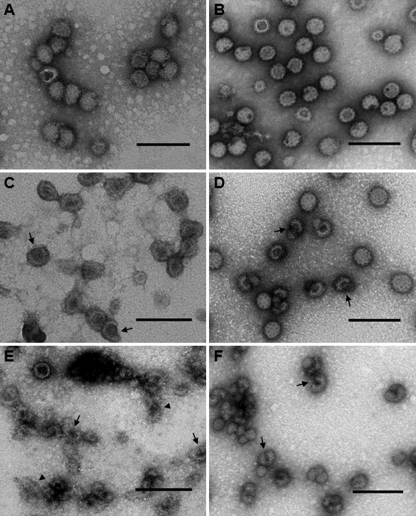
FIGURE 3.
Effect of cholesterol depletion on MAYV structure as assessed by negative-staining electron microscopy. HCCPs (A, C, and E) and LCCPs (B, D, and F) were treated with 0 (A and B), 10 (C and D), or 25 (E and F) mm MβCD, prepared for negative staining and then analyzed by transmission electron microscopy. Arrows in panels C–F point to breaks in the viral envelope from which nucleocapsids leaked into the surrounding environment, while arrowheads in panel E indicate probable membrane debris from severely damaged virus particles. Bars: 100 nm.

