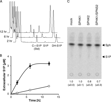
FIGURE 1.
hSPNS2-mediated release of endogenous S1P and DH-S1P. Shown are an HPLC diagram (A) and quantitative results (B) of time-dependent secretion of endogenous S1P and DH-S1P from cells. CHO cells expressing SPHK1 and hSPNS2 were incubated with the releasing medium for 2, 6, or 12 h. The releasing medium was collected, and the amounts of secreted S1P and DH-S1P were measured by HPLC. C17-S1P was used as the internal standard. Open and closed circles indicate secreted S1P and DH-S1P, respectively. Experiments were performed more than three times, and error bars indicate the S.D. C, sphingosine kinase activity in culture medium. The activity of SPHK was analyzed by the conversion of [3H]Sph to [3H]S1P. Lipid extracts were separated on a TLC plate, and bands corresponding to [3H]S1P were quantified. The relative amount of [3H]S1P in the medium of CHO/SPHK1 (SPHK1), CHO/SPHK2 (SPHK2), or CHO/SPHK1/hSPNS2 (SPHK1/SPNS2) compared with CHO (mock) cells is indicated at the bottom of the TLC plate image. Experiments were performed more than three times, and values are means with S.D.

