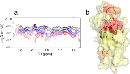
FIGURE 6.
a, two-dimensional diffusion-ordered spectroscopy map for β2-m in pure water in the presence of doxycycline at 1:0 molar ratio (black), at 1:1 molar ratio (blue), and at 1:3 molar ratio (red). The diffusion coefficients were estimated in the limit of a globular isotropic assumption for aqueous β2-m, using the trimeric EMILIN1 gC1q D value as a calibrator for a 51.2-kDa isotropic globule (41). b, β2-m interaction sites with doxycycline in pure water. These locations were obtained from the 15N-1H HSQC chemical shift (Δδ) and intensity change analysis upon doxycycline titration. The compensated Δδ values were calculated as described under “Experimental Procedures” after Mulder et al. (27). The color code is: yellow, the residues with Δδ ≤ Δδ + σ, where Δδ is the mean chemical shift change and σ is the chemical shift difference standard deviation; orange, residues with Δδ + σ < Δδ ≤ Δδ + 2σ; red, residues with Δδ > Δδ Δδ + 2σ.

