FIGURE 8.
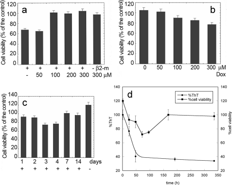
Effects of doxycycline on cytotoxicity of β2-m. a, β2-m (100 μm) was resolubilized in the absence or in the presence of different concentrations of doxycycline at 37 °C for 1.0 h. 10-μl aliquots of the samples were diluted in 90 μl of cell medium and added to wells coated with SH-SY5Y cells whose viability is reported as a function of the initial doxycycline concentration. b and c, preformed β2-m fibrils (100 μm, monomer concentration) were treated with doxycycline (Dox) for 72 h at the indicated concentrations (b) or at a fixed concentration (300 μm) for the indicated time lengths (c) and then added to the culture medium to estimate toxicity. Cell viability was measured by the MTT assay. The data are expressed as the percentage with respect to the control values obtained in three independent experiments, each value being the average of six trials. d, effect of the time of exposure to doxycycline of preformed β2-m fibrils on cell viability and ThT fluorescence quenching (same experiment as c). In all panels, error bars indicate means ± S.D.

