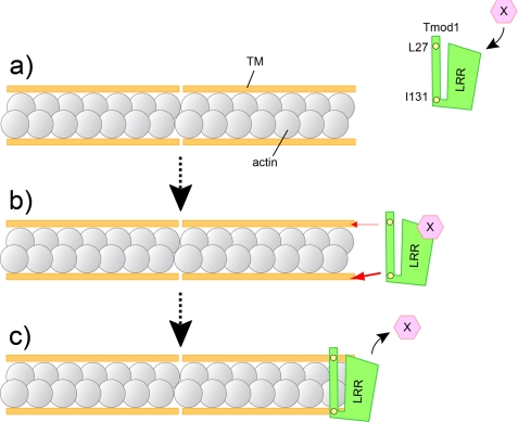
FIGURE 10.
Possible in vivo model of the Tmod1 capping at thin filament pointed ends in cardiomyocytes. a, an unidentified regulatory factor (X) binds to the LRR within Tmod1; the interaction assists Tmod1 to be targeted to thin filament pointed ends in cardiomyocytes. b, both TM-binding sites 1 and 2 are required for proper assembly of Tmod1 to the thin filament pointed ends in cardiac myocytes. Yellow circles are critical amino acid residues for TM binding site 1 (Leu-27) and site 2 (Ile-131), respectively. TM-binding site 2 contributes to the pointed end distribution of Tmod1 more than TM-binding site 1 (the thickness of the red arrow indicates the degree of contribution). The differences presumably guide Tmod1 to thin filament pointed ends with correct orientation. c, model for Tmod1 actin filament pointed end capping based on previous in vitro observation (17, 26) and present results. One Tmod1 molecule cooperatively binds two molecules of TM and interacts with at least one or two actin molecules at the pointed end.

