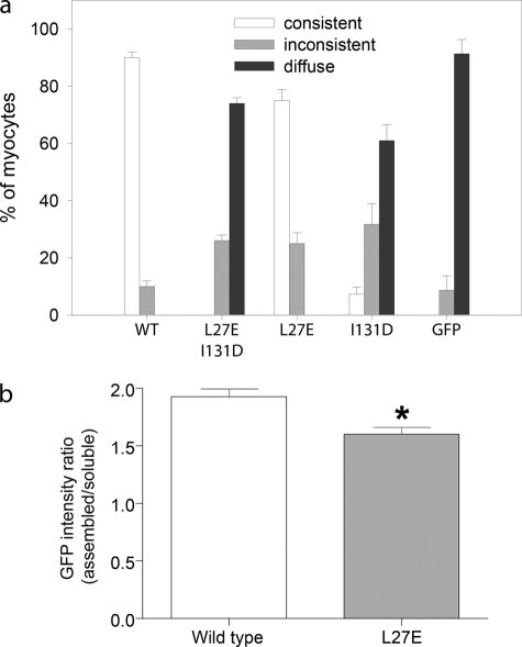
FIGURE 3.
Tropomyosin-binding site 2 is primarily responsible for determining the pointed end localization of Tmod1 in cardiomyocytes. a, graph shows the percentage of myocytes demonstrating consistent, inconsistent, or diffuse thin filament pointed end localization (mean ± S.D.); categories are as described under “Experimental Procedures.” A single mutation within tropomyosin-binding site 2 (I131D) strikingly perturbed Tmod1 pointed end localization (7.3 ± 2.5% cells displayed consistent localization versus 90.0 ± 2.0% consistent localization in cells expressing WT GFP-Tmod1). The localization was completely abolished when an additional mutation within another tropomyosin-binding site (L27E, site 1) was added. However, the single mutation within site 1 (L27E) did not have a strong effect on pointed end localization (75.0 ± 3.8% cells displayed consistent localization). b, graph shows the GFP intensity ratio (assembled/soluble GFP) determined by ImageJ software (Student's t test; *, p < 0.01). Cells expressing GFP-Tmod1(L27E) displayed more diffuse/soluble GFP staining in the cytoplasm compared with cells expressing WT GFP-Tmod1.

