FIGURE 4.
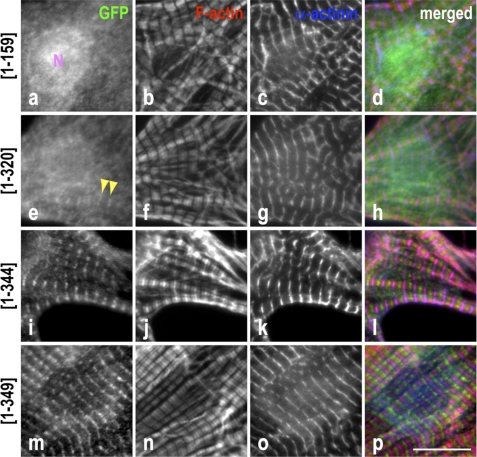
The LRR fold plus the C-terminal α-helix of Tmod1 are required for efficient localization to the pointed ends of thin filaments. Neonatal rat cardiomyocytes expressing GFP-Tmod1 fragments were stained for GFP (a, e, i, and m), F-actin using fluorescently conjugated phalloidin (b, f, j, and n), and α-actinin to stain the Z-discs (c, g, k, and o). d, h, l, and p show merged images of the triple staining (GFP, green; F-actin, red; α-actinin, blue). Clear and consistent striated pointed end localization of GFP-Tmod1(1–344) and GFP-Tmod1(1–349) (i and m) was observed, whereas GFP-Tmod1(1–159) (a) was diffuse in the cytoplasm with nuclear localization (N). Rare and faint (inconsistent) pointed end localization was observed for GFP-Tmod1(1–320) (e, arrowheads). Scale bar = 10 μm.

