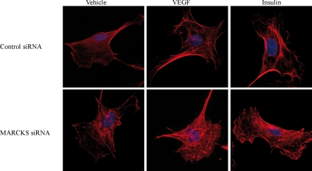
FIGURE 6.
Effects of siRNA-mediated MARCKS knockdown on the endothelial cell actin cytoskeleton. Shown are representative photomicrographs obtained in endothelial cells that were transfected with control or MARCKS siRNA, treated for 30 min with vehicle, VEGF (20 ng/ml), or insulin (100 nm) as shown, and then fixed and stained with phalloidin/Alexa Fluor 568 using the manufacturer's protocols. Fluorescent micrographic images were analyzed at λ = 573 nm using an Olympus DSU confocal imaging system (magnification ×100).

