Fig. 4.
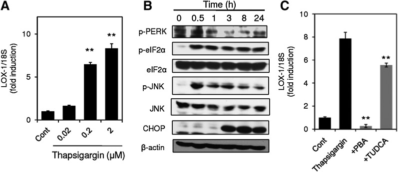
ER stress caused by thapsigargin upregulates LOX-1 expression. A: LOX-1 expression in THP-1 cells stimulated with thapsigargin for 24 h at the indicated concentrations was analyzed by real-time PCR. Data are expressed as means ± SE of three independent experiments. ** P < 0.01 versus Cont. B: THP-1 cells were incubated with 2 μM thapsigargin for the indicated periods, and cell lysates were subjected to Western blot analysis for phospho-PERK and β-actin. C: Effects of PBA and TUDCA on thapsigargin-induced LOX-1 expression. THP-1 cells were stimulated with 2 μM thapsigargin in the presence or absence of PBA and TUDCA for 24 h. LOX-1 expression was quantified by real-time PCR and normalized relative to 18S rRNA. Data are expressed as means ± SE of three independent experiments. ** P < 0.01 versus thapsigargin.

