FIG. 1.
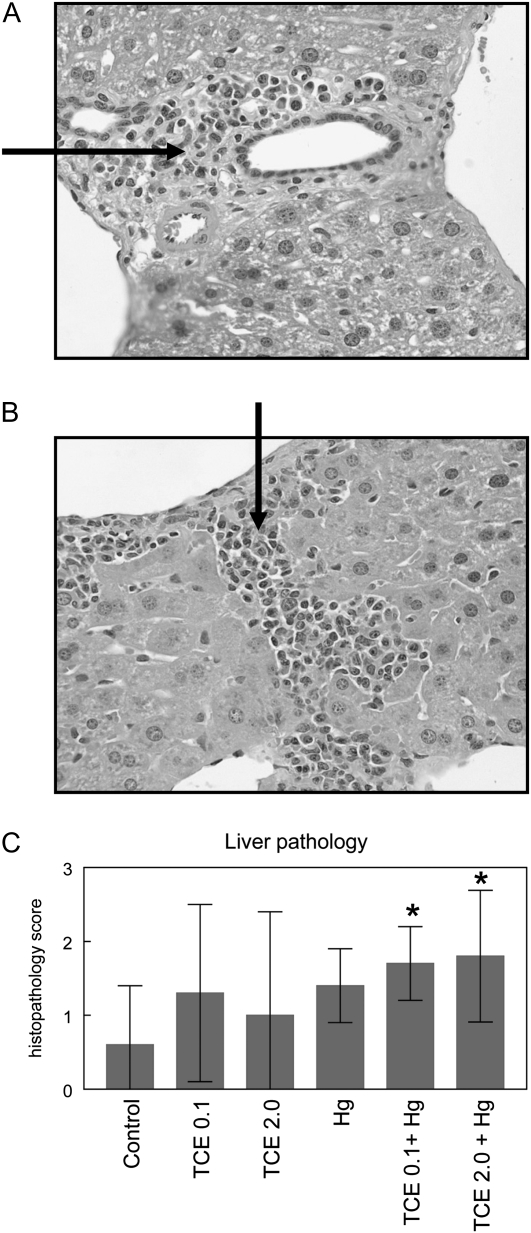
Coexposure to TCE and HgCl2 promoted AIH. Shown are representative liver sections (×400) demonstrating portal inflammation with fibrosis (A) and centrilobular inflammation (B) in liver sections from mice exposed to both TCE (2 mg/ml) and HgCl2. The arrows indicate areas of mononuclear infiltration. (C) Cumulative histopathology scores for liver sections. *Statistically different from the results obtained using liver sections from control mice.

