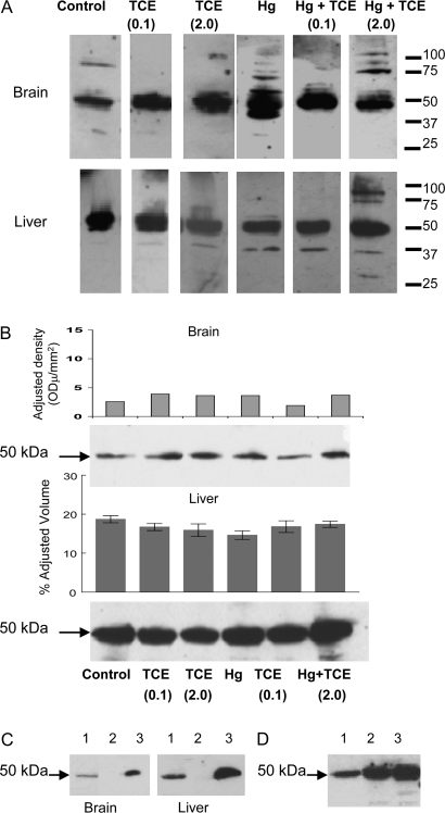
FIG. 7.
Detecting TCE- and HgCl2-induced antibodies after 4 week exposure. (A) Pooled microsomal liver or brain protein collected from control MRL+/+ mice (treated with water alone for 8 weeks) was separated by SDS-PAGE and immunoblotted using pooled sera from pooled control or treated MRL+/+ mice collected at the 4-week time point, followed by a goat anti-mouse IgG antibody. (B) Brain or microsomal liver proteins from control MRL+/+ mice or MRL+/+ mice exposed to either TCE and/or HgCl2 for 8 weeks were separated by SDS-PAGE and immunoblotted with goat anti-mouse IgG antibody alone. Densitometric analysis of the 50-kDa protein was conducted. This experiment was repeated with brain tissue, and mean densitometric values (± SD) from three experiments are presented. (C) Pooled brain or microsomal liver protein from control MRL+/+ mice was separated by SDS-PAGE and immunoblotted with goat anti-mouse IgG antibody (lane 1). Alternatively, IgG was immunoprecipitated from the protein lysates using beads conjugated with donkey anti-mouse IgG; the remaining lysates (lanes 2) or resulting beads (lanes 3) were separated by SDS-PAGE and immunoblotted with goat anti-mouse IgG. (D) Microsomal liver protein from an untreated MRL+/+ mouse (lane 1), C57BL/6 mouse (lane 2), or C3H/HeJ mouse (lane 3) was separated by SDS-PAGE and immunoblotted with goat anti-mouse IgG antibody.

