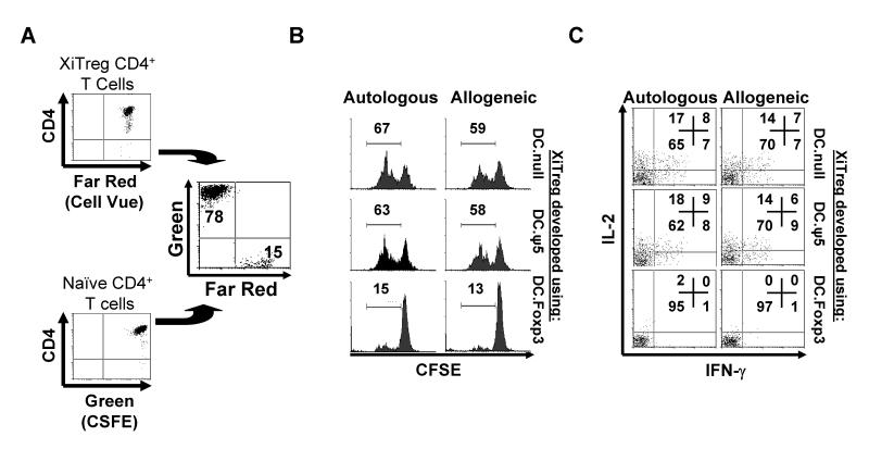
Figure 6. DC.Foxp3 (but not control DC)-induced XiTreg suppress the proliferation of, and Type-1 cytokine (IFN-γ and IL-2) production by, CD4+ T cells in vitro.
A, XiTreg were generated in 21 day cultures as described in the legend of Fig. 5 and labeled with Cell Vue Claret stain per the manufacturer’s protocol. Cell Vue Claret stained-XiTreg and CFSE-labeled CD4+CD45ROnegCD25neg T cells were then added to wells at a 10:1 responder-to-XiTreg ratio (A), along with anti-CD3/CD28 microbeads. After 72h of coculture, the Cell Vue-excluded population of CD4+ T cells was analyzed for proliferation (i.e. CFSE dilution; panel B) and for intracellular levels of IL-2 and IFN-γ (panel C) by flow cytometry. For each panel, similar data were obtained in 3 independent experiments. Transduction efficiency for DC.Foxp3 used to generate XiTreg: 51% .

