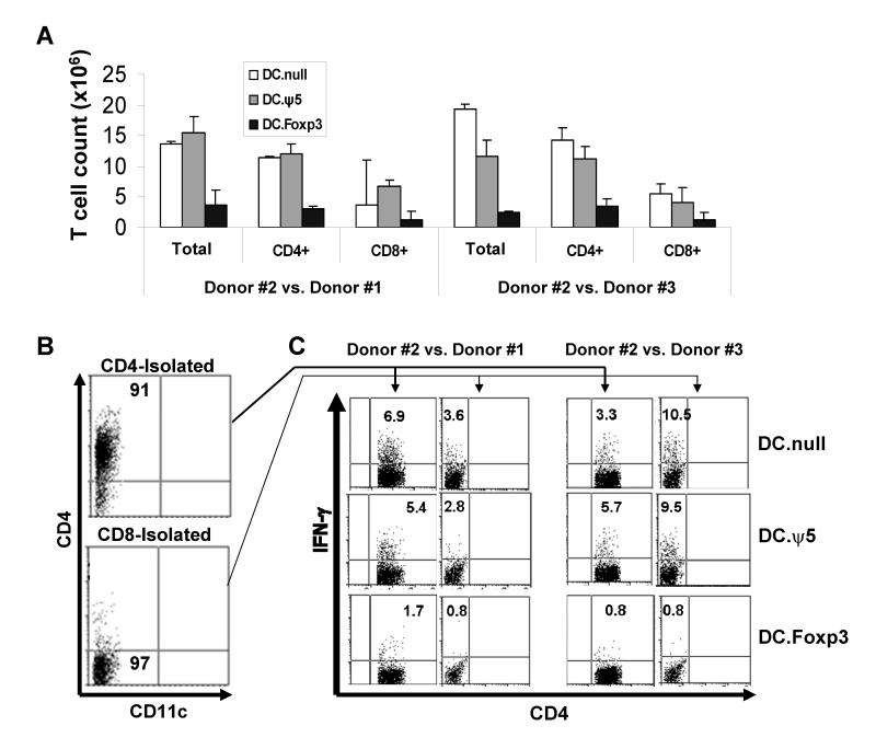
Figure 9. Alloreactive CD4+ and CD8+ T cells primed by DC.Foxp3 expand poorly and fail to exhibit Type-1 responses against recall or third-party alloantigens.
DC.Foxp3 or control DC were generated from normal donor #1 and cultured with total CD45ROneg T cells isolated from unrelated, normal donor #2 at a DC:T cell ratio of 1:10. In panel A, day 7 cultures were analyzed for T cell yields. Purified CD4+ or CD8+ T cells harvested from these cultures (panel B) were (re)stimulated with DC.null generated either from normal donor #1 or a third-party, normal donor #3 in the presence of rhIL-2 and rhIL-7. After 7 additional days of culture, CD4+ and CD8+ (i.e. depicted as CD4neg) T cells were analyzed for intracellular levels of IFN-γ by flow cytometry (panel C). For each panel, similar data were obtained in 3 independent experiments. Transduction efficiency for DC.Foxp3: 53% in panels A-C.

